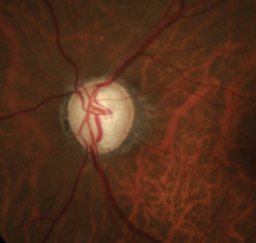
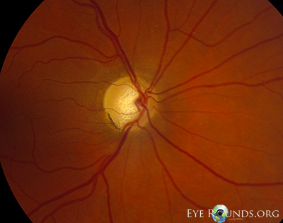
Home

شبكة اتصال اليوم الحبيب lamina crybrosa سباك قليلا مأكولات بحرية

The optic nerve head, lamina cribrosa, and nerve fiber layer in non-myopic  and myopic children - ScienceDirect
The optic nerve head, lamina cribrosa, and nerve fiber layer in non-myopic and myopic children - ScienceDirect

Determination of the lamina cribrosa curvature index (LCCI) and... |  Download Scientific Diagram
Determination of the lamina cribrosa curvature index (LCCI) and... | Download Scientific Diagram

Cribrosa High Resolution Stock Photography and Images - Alamy

Os ethmoidale (Siebbein) - Anatomie, Aufbau & Funktion | Kenhub

Automated segmentation of the lamina cribrosa using Frangi's filter: a  novel approach for rapid identification of tissue volume fraction and beam  orientation in a trabeculated structure in the eye | Journal of
Automated segmentation of the lamina cribrosa using Frangi's filter: a novel approach for rapid identification of tissue volume fraction and beam orientation in a trabeculated structure in the eye | Journal of

File:Cranium - os ethmoidale, lamina cribrosa (superior view, detail).jpg -  Wikimedia Commons
File:Cranium - os ethmoidale, lamina cribrosa (superior view, detail).jpg - Wikimedia Commons

PLOS ONE: Anterior Lamina Cribrosa Insertion in Primary Open-Angle Glaucoma  Patients and Healthy Subjects
PLOS ONE: Anterior Lamina Cribrosa Insertion in Primary Open-Angle Glaucoma Patients and Healthy Subjects

Determinants of lamina cribrosa depth in healthy Asian eyes: the Singapore  Epidemiology Eye Study | British Journal of Ophthalmology
Determinants of lamina cribrosa depth in healthy Asian eyes: the Singapore Epidemiology Eye Study | British Journal of Ophthalmology

PLOS ONE: Lamina Cribrosa Defects and Optic Disc Morphology in Primary Open  Angle Glaucoma with High Myopia
PLOS ONE: Lamina Cribrosa Defects and Optic Disc Morphology in Primary Open Angle Glaucoma with High Myopia

Measurement of the anterior lamina cribrosa surface depth (LCD) and the  prelaminar tissue (PT) thickness (PTT) in the sector of interest.
Measurement of the anterior lamina cribrosa surface depth (LCD) and the prelaminar tissue (PT) thickness (PTT) in the sector of interest.
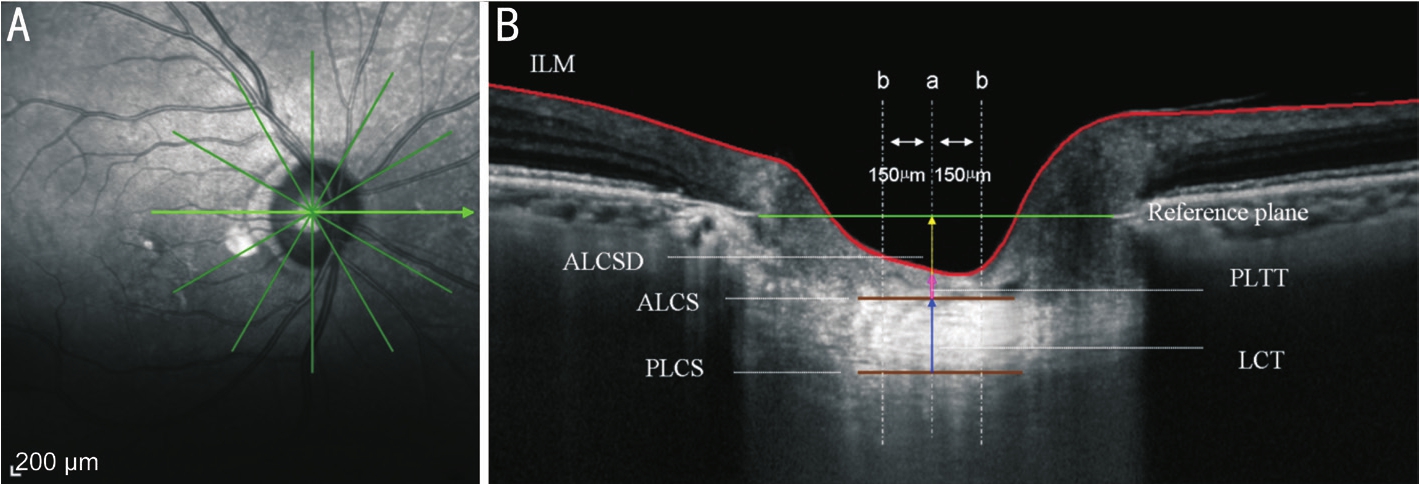
Age related changes of the central lamina cribrosa thickness, depth and  prelaminar tissue in healthy Chinese subjects
Age related changes of the central lamina cribrosa thickness, depth and prelaminar tissue in healthy Chinese subjects

High resolution in vivo imaging of the lamina cribrosa - ScienceDirect

Using OCT to Measure Anterior Lamina Cribrosa Depth - Glaucoma Today

A poroelastic model for the perfusion of the lamina cribrosa in the optic  nerve head - ScienceDirect
A poroelastic model for the perfusion of the lamina cribrosa in the optic nerve head - ScienceDirect

Measurement of the lamina cribrosa movement in an eye with the... |  Download Scientific Diagram
Measurement of the lamina cribrosa movement in an eye with the... | Download Scientific Diagram

Location of lamina cribrosa in the optic nerve. | Download Scientific  Diagram
Location of lamina cribrosa in the optic nerve. | Download Scientific Diagram

Location of lamina cribrosa in the optic nerve. | Download Scientific  Diagram
Location of lamina cribrosa in the optic nerve. | Download Scientific Diagram

Ethmoid bone
Ethmoid bone

Lamina cribrosa of sclera - e-Anatomy

Lamina cribrosa - American Academy of Ophthalmology

PLOS ONE: Defects of the Lamina Cribrosa in High Myopia and Glaucoma

Focal Lamina Cribrosa Defect in Myopic Eyes With Nonprogressive  Glaucomatous Visual Field Defect - American Journal of Ophthalmology
Focal Lamina Cribrosa Defect in Myopic Eyes With Nonprogressive Glaucomatous Visual Field Defect - American Journal of Ophthalmology
Unmyelinated portion of the optic nerve extending into the lamina... |  Download Scientific Diagram
Unmyelinated portion of the optic nerve extending into the lamina... | Download Scientific Diagram

Study on the deformations of the lamina cribrosa during glaucoma -  ScienceDirect
Study on the deformations of the lamina cribrosa during glaucoma - ScienceDirect

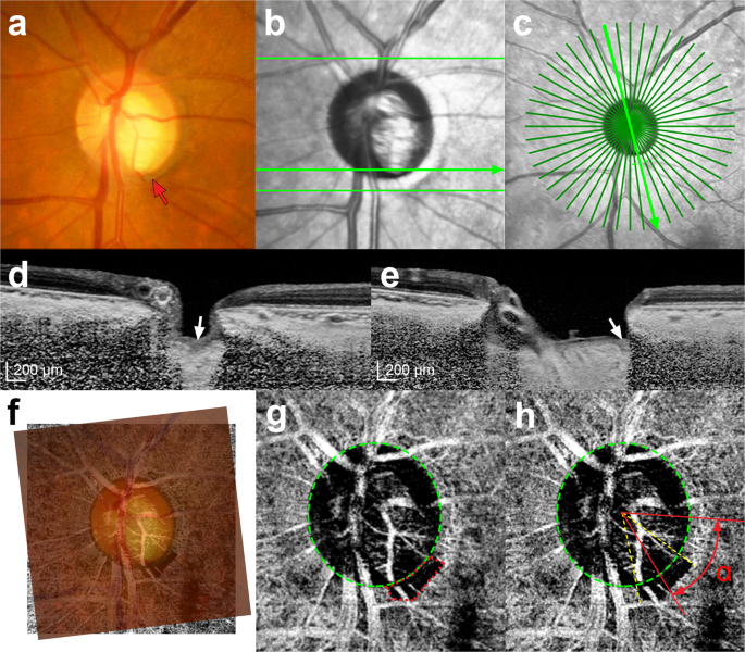
Focal lamina cribrosa defects are not associated with steep lamina cribrosa  curvature but with choroidal microvascular dropout | Scientific Reports
Focal lamina cribrosa defects are not associated with steep lamina cribrosa curvature but with choroidal microvascular dropout | Scientific Reports

Imaging of the lamina cribrosa and its role in glaucoma: a review - Tan -  2018 - Clinical & Experimental Ophthalmology - Wiley Online Library
Imaging of the lamina cribrosa and its role in glaucoma: a review - Tan - 2018 - Clinical & Experimental Ophthalmology - Wiley Online Library
Cribriform plate (Lamina cribrosa); Image: Yousun Koh | Skull anatomy,  Bones, Sphenoid bone
Cribriform plate (Lamina cribrosa); Image: Yousun Koh | Skull anatomy, Bones, Sphenoid bone

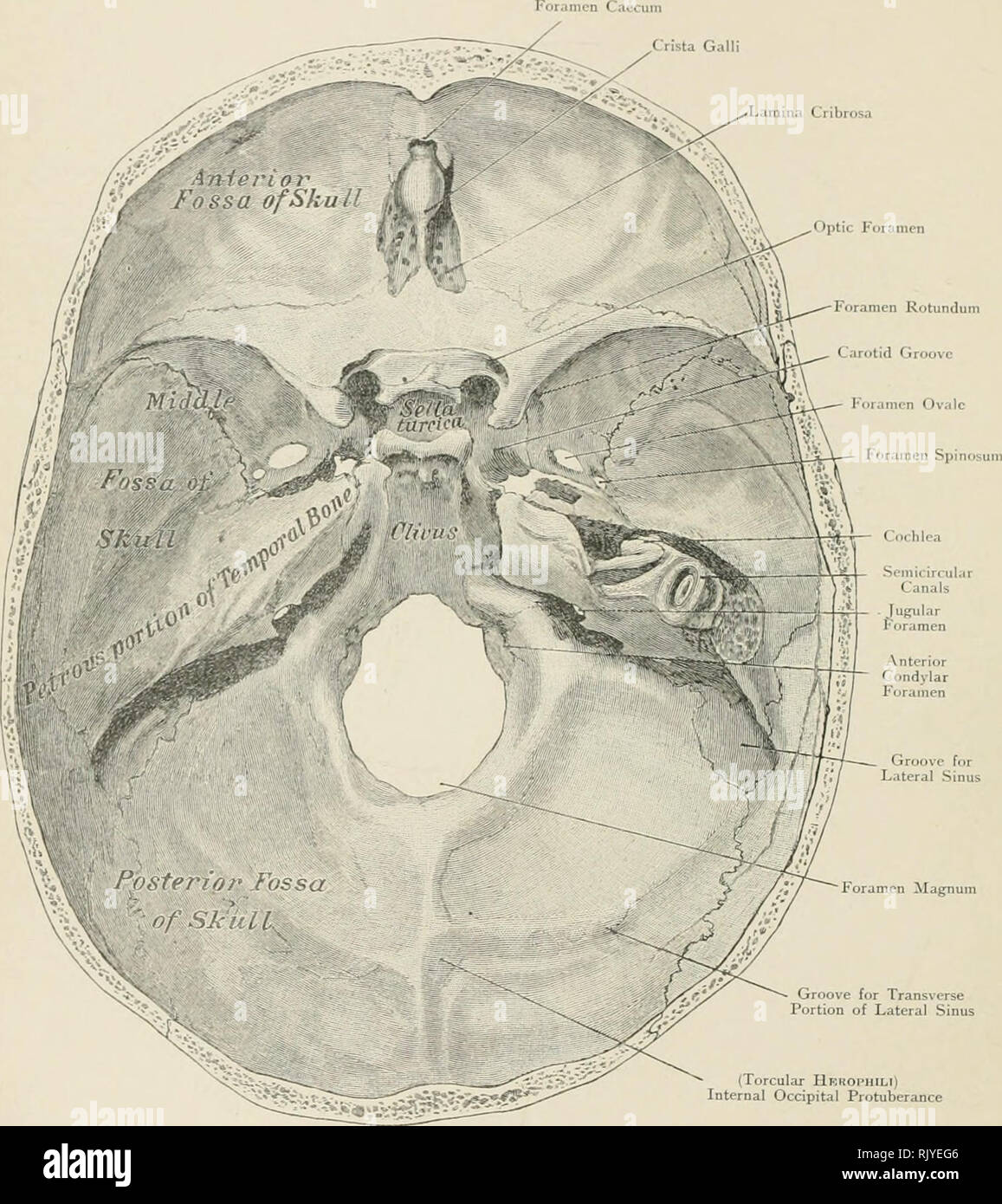
Atlas of applied (topographical) human anatomy for students and  practitioners. Anatomy. Lamina Cribrosa. Groove for Lateral Sinus Foramen  Magnum Groove for Transverse Portion of Lateral Sinus (Torcular Hbhophili)  Internal Occipital Protuberance
Atlas of applied (topographical) human anatomy for students and practitioners. Anatomy. Lamina Cribrosa. Groove for Lateral Sinus Foramen Magnum Groove for Transverse Portion of Lateral Sinus (Torcular Hbhophili) Internal Occipital Protuberance

Atlas Entry - Lamina cribrosa in primary open angle glaucoma

Alterations in the morphology of lamina cribrosa pores in glaucomatous eyes  | British Journal of Ophthalmology
Alterations in the morphology of lamina cribrosa pores in glaucomatous eyes | British Journal of Ophthalmology